Hey everyone, let’s cut to the chase—if you’ve ever wondered what actually happens when someone’s having a heart attack, and how we spot it with medical imaging, this post’s for you. I’m not here to give a stuffy textbook lecture, promise—just a heads-up from an imaging supplier who’s spent years working with clinics and hospitals on tools that catch these things fast. Let’s start with the basics: a heart attack isn’t just “chest pain”—it’s when blood flow to part of the heart muscle gets blocked, usually by a blood clot sitting on top of a cholesterol plaque that’s built up in a coronary artery. The faster we spot that blockage, the less damage the heart takes. That’s where imaging comes in, and it’s way more varied than just an EKG (though we love EKGs too—don’t sleep on those). Imaging
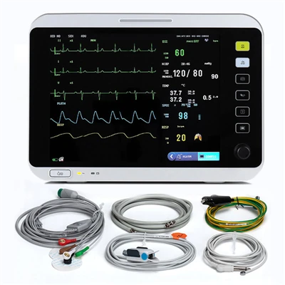
First up, let’s talk about the go-to first tests in the ER: the 12-lead EKG. I know, EKGs are like the old reliables, but hear me out—they’re imaging in their own weird way. What you’re actually seeing on that paper strip is the electrical activity of the heart. When part of the heart muscle is starved of oxygen (which is exactly what happens during a heart attack), the electrical signals go haywire. For example, ST-elevation myocardial infarction (STEMI) is the big one we look for—those “elevated ST segments” on the EKG line that pop up in certain leads. That’s a pretty clear sign of a full blockage, and it means doctors need to act fast. The non-STEMI (NSTEMI) ones are trickier—those ST segments get depressed, or T waves flip, which can be easy to mix up with other things like anxiety or electrolyte issues. EKGs are super quick (takes like 10 seconds to do) which is why they’re the first line, but they’re not the whole story. That’s when we bring in the fancier imaging.
Next, let’s chat about echocardiograms—aka heart ultrasounds. These are the ones with the probe that moves over your chest, like a regular ultrasound for a baby but way more high-stakes. What do they show during a heart attack? The motion of the heart wall. Normally, all the segments of your heart squeeze in sync, right? But if a part of the muscle just got oxygen cut off, that segment stops moving, or moves weirdly—like it bulges out when the rest of the heart squeezes. We call that “wall motion abnormality,” and it shows up within minutes of a heart attack, even before the EKG changes sometimes. Another thing echocardiograms pick up are things like heart valve issues (though that’s less acute for heart attacks) or if blood’s leaking where it shouldn’t. It’s also great for checking the ejection fraction—how well the heart is pumping blood overall. I’ve seen cases where a patient came in with vague chest pain, EKG was weird, but an echocardiogram spotted a huge wall motion problem that locked in the heart attack diagnosis. Game-changer for ruling out other stuff like pericarditis (that’s inflammation around the heart, not the muscle itself).
Then there’s cardiac CT—computed tomography. This one’s gotten way better over the last 10 years, with those fast CT scanners that can snap a full set of heart images in like 5 seconds. Wait, why would you use CT for a heart attack? Well, if someone comes in with chest pain and we’re not sure if it’s a heart attack, CT can rule out other causes fast—like a pulmonary embolism (a clot in the lung) or aortic dissection (a tear in the big artery coming off the heart, which is life-threatening too). But we also use CT to look directly at the coronary arteries. Wait, can CT see the plaque? Yep—soft plaque, the kind that’s prone to rupturing (the stuff that causes heart attacks) shows up darker than the hard, calcified plaque. And if there’s a big blockage, you can see the contrast dye we inject not flowing through that spot. The catch is, you have to hold your breath for a few seconds, and it uses a bit more radiation than other tests, but it’s super useful for intermediate cases where EKG and echocardiogram are ambiguous. We’ve worked with a lot of hospitals that use cardiac CT for “rule out heart attack” patients because it cuts down on stay times so much—patients don’t have to wait 6 hours for enzyme tests, they get a clear answer fast.
Now, let’s not leave out cardiac MRI—this is the fancy one, the gold standard for a lot of post-heart attack checks, but sometimes used acutely too. What makes MRI different? It uses magnets and radio waves, no radiation, and it can super detailed look at the heart muscle itself. During a heart attack, the damaged muscle (the “infarct”) shows up bright on certain sequences—like late gadolinium enhancement, where the contrast dye gets trapped in the dead tissue. This is great for figuring out how old the heart attack is (was it hours ago, days ago, weeks ago?) and how big the damage is. It can also tell if there’s any damage to the heart valves or the lining, or if the patient had something like a viral infection instead of a heart attack. The only downsides are that it’s slower (takes 20-30 minutes, sometimes longer) and you can’t have certain metal implants, so it’s not great for super unstable patients in the ER. But for follow-up, it’s unbeatable.
Wait, let’s talk about what the lab tests (like troponin) have to do with imaging, because people often mix them up. Troponin is a protein that leaks out of damaged heart muscle, right? It takes a few hours after the heart attack starts to show up in the blood, so imaging often spots the problem before the blood test comes back. That’s why imaging is so critical for fast treatment—you don’t want to wait 6 hours for troponin if the patient is having a STEMI, you need to get them to the cath lab right away. I’ve heard ER docs say time is muscle—every minute the blockage is there, more heart tissue dies. So having imaging that gives a quick answer is huge.
Now, let’s get real about what you don’t see. A lot of people think imaging can see the actual clot, and sometimes we can with coronary angiography (that’s the one where they thread a catheter up to the heart arteries and inject dye, then take X-rays). That’s the most invasive test, usually done in the cath lab right when a STEMI is diagnosed. It shows exactly where the blockage is, so doctors can put in a stent to open the artery. But coronary angiograms only show the lumen (the inside of the artery) — you can see that it’s blocked, but you don’t get the whole picture of the plaque. That’s where CT and intravascular ultrasound (IVUS) come in—IVUS is like a tiny ultrasound probe on a catheter, so you can see the inside of the artery wall, the plaque type, how big it is, if it’s ruptured. That’s super useful for patients who might need more than a stent, or for cases where the angiogram looks weird but it’s not clear if it’s a blockage or something else.
Wait, let’s throw in a real-world example to make this stick. Last year, we worked with a small community hospital that was getting a lot of patients coming in with chest pain that turned out not to be heart attacks. They had a old EKG machine that was finicky, and their echocardiogram tech was new, so they were missing a lot of cases. We helped them upgrade their echocardiogram systems and train their team on how to spot subtle wall motion abnormalities. Within 6 months, their time to diagnosis for STEMI patients dropped by 25%, and they reduced the number of unnecessary cath labs activations by 18%—that’s a win for everyone, right? Patients got treated faster, and the hospital saved money on tests they didn’t need.
I know there’s a lot of info here, but the key takeaway is this: heart attack imaging isn’t one-size-fits-all. It starts with a quick EKG to rule out STEMI immediately, then uses echocardiogram for real-time heart function, cardiac CT or MRI for ambiguous cases or follow-up, and invasive angiography with IVUS to get the exact details for treatment. As an imaging supplier, we’re all about making these tools accessible, easy to use, and accurate—because at the end of the day, every second counts when someone’s heart is in trouble.
Diabetes Healthcare If you’re a clinic, hospital, or healthcare provider looking to upgrade your imaging systems, streamline your heart attack diagnosis workflow, or just have questions about how these tools work, reach out to us. We partner with teams of all sizes, from small community hospitals to big academic medical centers, to make sure they have the right tools to catch heart attacks fast and save lives. Don’t hesitate to connect with our team to talk through your specific needs—we’re here to help.
References
- American Heart Association. (2023). "Coronary Heart Disease: Heart Attack Imaging." AHA.org.
- Braunwald, E. (2022). Braunwald’s Heart Disease: A Textbook of Cardiovascular Medicine (12th ed.). Elsevier.
- American College of Cardiology. (2021). "Clinical Guidelines for the Evaluation and Management of Chest Pain." ACC.org.
- Hilfiker, P., et al. (2020). "Cardiac Computed Tomography for Acute Chest Pain: A Systematic Review." Journal of the American College of Cardiology: Cardiovascular Imaging.
Eurosun Technology (Shanghai) Co., Ltd.
As one of the most professional imaging equipment manufacturers and suppliers in China, we have world-leading production equipment and strong manufacturing capabilities. We warmly welcome you to wholesale high quality imaging equipment in stock here from our factory.
Address: No.485, Xingmei Road, Minhang District, Shanghai
E-mail: eurosun6661216@163.com
WebSite: https://www.eurosunmedical.com/
